- Name of the project: Implementation of the research Nr.1.10. „Research on the reconstruction technology of the volumetric three-dimensional images” – 3DAtA
- Partners: EUROLCDS and “Hanza Elektronika”
- Aim of the project: Medical systems that using nuclear magnetic resonance, X-ray and other methods to acquire spatial exploration images of patient, stores them in a medical image standard – DICOM. In this standard, the 3D images are stored on “cut” layers and stored, packaged in various service information. In this research there are develope optimum DICOM image transformation methods that convert spatial 3D images in the form of a matrix that would allow them to output to a 3D three-dimensional image display.
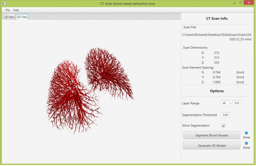
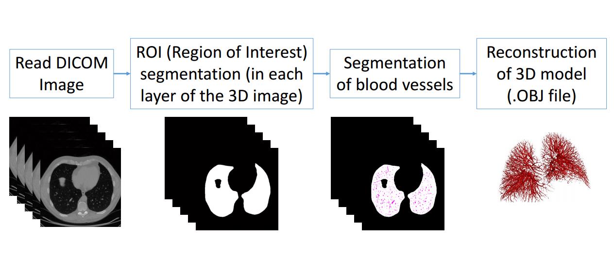
Developed technologies: In this project software for automatic blood vessel segmentation from Lung Computed Tomography scans and 3D model construction was developed. Publicly available CT scan database was used for algorithm development: https://vessel12.grand-challenge.org. Automatic blood vesel segmentation was based on Stacked Multiscale Feature Learning methods. 3D model was generated using marching cubes algorithm. For 3D model generation software with graphical user interface was developed. Software with developed using Java framework. 3D model can be saved in wafefront format, which can be used for vizualisation on volumetric 3D display.
Results achieved: Development of method and software, that allows to segment lung blood vessels and construct 3D model in waveftont format, for vizualization in volumertric 3D display.